Transanal minimally invasive surgery (TAMIS) is designed to enable surgeons to remove rectal tumours and polyps through the anus without external abdominal incisions. This technique provides access to lesions located 4-15cm from the anal verge, reaching areas that may be difficult to access with traditional transanal excision. For patients with early-stage rectal cancers or large benign polyps, transanal minimally invasive surgery in Singapore offers a potential sphincter-preserving option compared to more extensive operations.
TAMIS evolved from transanal endoscopic microsurgery (TEM), which required specialised equipment and a steep learning curve. The TAMIS approach uses single-incision laparoscopic ports that most colorectal surgeons already have experience with, making the technique more accessible while maintaining comparable clinical outcomes. The procedure typically takes 60-90 minutes and many patients return home within 24-48 hours.
How TAMIS Differs from Traditional Rectal Surgery
Traditional transanal excision uses retractors to visualise and remove lesions near the anal opening, but this approach becomes increasingly difficult for tumours higher in the rectum. Surgeons may lose direct visualisation, making precise excision margins more challenging to achieve.
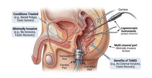
Transanal minimally invasive surgery aims to address these limitations through insufflation, filling the rectum with carbon dioxide gas to create a working space. A single-port device inserted through the anus provides channels for a camera and laparoscopic instruments. This setup gives surgeons magnified views and precise instrument control throughout the rectum.
The full-thickness excision capability distinguishes TAMIS from endoscopic polypectomy. While colonoscopic removal works well for polyps confined to the mucosal layer, TAMIS excises all layers of the rectal wall. This matters for staging purposes and helps achieve adequate margins around suspicious lesions.
Compared to low anterior resection, where the affected rectum segment is removed and the bowel reconnected, TAMIS is designed to preserve rectal anatomy. Patients typically maintain normal bowel function without requiring the temporary or permanent stoma that major rectal surgery may involve.
Conditions Treated with TAMIS
Early-Stage Rectal Cancer (T1)
Rectal cancers confined to the submucosa (T1) without lymph node involvement may qualify for TAMIS as definitive treatment. Tumours must be well or moderately differentiated, without lymphovascular invasion, and smaller than 3cm. Pre-operative MRI and endorectal ultrasound determine the depth of invasion and help identify suitable candidates.
TAMIS provides an intact specimen for detailed pathological analysis. If the final pathology reveals unfavourable features, such as deeper invasion than expected, positive margins, or high-risk characteristics, the patient can proceed to radical surgery while still in the early post-operative window.
Large Rectal Polyps
Polyps too large or positioned awkwardly for safe colonoscopic removal represent common TAMIS indications. Sessile polyps spreading across multiple rectal folds and those with concerning features on biopsy benefit from full-thickness excision. This approach reduces the perforation risk associated with aggressive endoscopic techniques for difficult lesions.
Rectal Neuroendocrine Tumours
Small rectal carcinoids without evidence of metastasis may respond to local excision. TAMIS aims to achieve clear margins while preserving rectal function.
Anastomotic Strictures and Fistulas
Beyond tumour excision, TAMIS provides access for repairing complications from previous rectal surgery. Surgeons can directly visualise and treat anastomotic strictures or small rectourinary fistulas through the TAMIS platform.
Who Qualifies for TAMIS
Ideal Candidates
Patients with lesions located 4-15cm from the anal verge may benefit from the TAMIS approach. Tumours must be smaller than 4-5cm to allow adequate visualisation and manipulation within the rectal lumen. Pre-operative imaging should show no evidence of lymph node involvement or distant metastasis for malignant lesions.
Medical fitness for general anaesthesia is required, though TAMIS places less physiological stress than major abdominal surgery. Patients with significant cardiopulmonary disease may tolerate TAMIS when they would not safely undergo low anterior resection.
Relative Contraindications
Previous pelvic radiation can distort tissue planes and impair healing, making TAMIS technically challenging. Prior rectal surgery may create adhesions that limit port placement. Anal stenosis or sphincter abnormalities may prevent adequate port insertion.
Circumferential or near-circumferential tumours risk stricture formation if excised via TAMIS. These lesions typically require radical resection to avoid significant bowel function impairment.
When TAMIS Isn’t Appropriate
T2 or deeper rectal cancers require radical surgery with lymphadenectomy, since the oncological benefit of removing potentially involved lymph nodes outweighs the functional advantages of TAMIS. Patients with confirmed nodal metastasis on imaging need formal resection regardless of the primary tumour’s size.
Lesions extending to the anal canal fall outside the TAMIS working space. Very low tumours may be accessible through traditional transanal excision without requiring the TAMIS platform.
The TAMIS Procedure: What Happens
Pre-Operative Preparation
Bowel preparation the day before surgery clears the rectum for adequate visualisation. Most surgeons prescribe oral laxatives rather than full mechanical bowel prep. Prophylactic antibiotics are administered before the procedure begins.
Patients undergo either general anaesthesia or spinal anaesthesia depending on the expected duration and tumour location. Positioning varies (such as lithotomy, prone jackknife, or lateral) based on where the lesion sits within the rectum.
During Surgery
The surgeon inserts a gel port or SILS port through the anal canal and establishes pneumorectum with carbon dioxide. A 5mm laparoscope provides the visual field while specialised instruments perform the dissection.
Marking the excision boundary with electrocautery helps achieve adequate margins around the lesion. The surgeon then incises through all rectal wall layers and develops the dissection plane in the mesorectal fat. For posterior tumours, the presacral space becomes visible; anterior lesions approach the prostate or vagina, requiring careful attention to these structures.
After removing the specimen, the defect is closed with absorbable sutures. Some surgeons leave small defects to heal by secondary intention, though closure appears to reduce complications.
Immediate Post-Operative Period
Patients typically experience mild rectal discomfort controlled with oral analgesics. Diet advances from clear liquids to regular food within hours of surgery. Early ambulation begins the same day.
Hospital stays average one to two nights. Some centres perform TAMIS as day surgery for straightforward cases, with patients discharged once they demonstrate stable vital signs and adequate oral intake.
💡 Did You Know?
The TAMIS platform allows surgeons to work through a natural body opening while achieving precision comparable to abdominal laparoscopic surgery. The camera magnifies the surgical field, revealing tissue planes that may not be visible to the naked eye.
Recovery and Expected Outcomes
The first week involves managing minor rectal bleeding, mucous discharge, and the sensation of rectal fullness. These symptoms resolve as the excision site heals. Many patients return to desk work within one week and physical occupations within two to three weeks.
Bowel function typically normalises within the first month. Some patients notice increased frequency or urgency temporarily, particularly after large excisions. These symptoms improve as the rectum regains compliance.
Follow-up appointments occur at two weeks for wound assessment and at six to eight weeks once pathology results guide further management. For benign polyps, surveillance colonoscopy follows standard intervals. Malignant lesions require closer monitoring with MRI, endoscopy, and clinical examination.
⚠️ Important Note
If pathology reveals cancer extending deeper than T1 or shows high-risk features, your surgeon will discuss whether additional treatment, potentially including radical surgery, is recommended. TAMIS does not prevent subsequent operations if needed.
Potential Complications
Bleeding
Minor bleeding occurs commonly and resolves spontaneously. Significant haemorrhage requiring intervention happens infrequently but may necessitate examination under anaesthesia with suturing or cauterisation.
Rectal Perforation
Full-thickness excision inherently creates a controlled perforation that surgeons close primarily. Uncontrolled perforation into the peritoneal cavity, more likely with anterior tumours, may require conversion to abdominal surgery for repair.
Urinary Retention
Spinal anaesthesia and rectal manipulation can temporarily affect bladder function. Most patients void normally after catheter removal, though some require short-term catheterisation.
Stricture Formation
Large excisions, particularly those extending more than half the rectal circumference, risk narrowing during healing. Strictures may require dilation or further surgery if symptomatic.
Local Recurrence
For malignant lesions, local recurrence rates depend heavily on proper patient selection. T1 cancers without adverse pathological features have a low local recurrence rate, comparable to radical surgery for appropriately selected tumours.
Comparing TAMIS to Other Approaches
| Approach | Access | Recovery | Bowel Function | Specimen Quality |
| Colonoscopic polypectomy | Through colonoscope | Same day | Typically unchanged | Piecemeal possible |
| Traditional transanal | Direct visualisation | 1-2 days | Typically unchanged | Limited to low rectum |
| TAMIS | Single port with insufflation | 1-2 days | Typically unchanged | Full-thickness, intact |
| Low anterior resection | Abdominal incisions | 5-7 days | Often altered | En bloc with lymph nodes |
| Abdominoperineal resection | Abdominal and perineal | 7-10 days | Permanent stoma | En bloc with lymph nodes |
✅ Quick Tip
Ask your surgeon whether your lesion’s location, size, and biopsy results make you a candidate for TAMIS. Bringing your colonoscopy report and any imaging to your consultation helps facilitate this discussion.
Questions to Ask Your Surgeon
Preparing specific questions helps maximise your consultation time. Consider asking about the surgeon’s experience with TAMIS, expected pathology turnaround time, and what happens if the pathology shows unexpected findings. Understanding the surveillance schedule after surgery and warning signs requiring urgent contact ensures you’re prepared for the recovery period.
When to Seek Professional Help
- Increasing pain: Increasing rectal pain not controlled with prescribed medications.
- Heavy bleeding: Heavy bleeding soaking through pads within hours.
- Fever: Fever above 38°C developing after discharge.
- Urinary retention: Inability to pass urine for more than eight hours.
- New incontinence: Faecal incontinence not present before surgery.
- Persistent nausea: Persistent nausea or vomiting preventing oral intake.
Commonly Asked Questions
How long does TAMIS surgery take?
TAMIS procedures typically take 60-90 minutes. Complex cases involving larger lesions or difficult locations may extend to two hours. The time includes port placement, excision, specimen retrieval, and defect closure.
Will I need a stoma bag after TAMIS?
TAMIS is designed to preserve rectal tissue, so a stoma is typically not required. This approach aims to maintain normal bowel continuity. If subsequent radical surgery becomes necessary based on pathology, stoma requirements would be discussed at that time.
When can I return to normal activities after transanal minimally invasive surgery?
Desk work typically resumes within one week. Driving is appropriate once you’re off narcotic pain medications and can perform emergency braking, usually within one to two weeks. Heavy lifting and strenuous exercise should wait four to six weeks.
Is TAMIS painful?
Many patients describe mild to moderate rectal discomfort rather than severe pain. Oral analgesics control symptoms for many patients. The absence of abdominal incisions avoids the wound pain associated with traditional surgery.
What if cancer is found unexpectedly?
If a polyp presumed benign contains cancer on final pathology, your surgical team reviews the specimen’s features. Low-risk cancers may need only surveillance. High-risk findings prompt discussion about additional surgery within a few weeks of the initial procedure.
Next Steps
TAMIS applies to rectal tumours and polyps that cannot be safely removed colonoscopically but do not require major abdominal surgery. Suitable candidates are those with lesions 4-15cm from the anal verge, confirmed T1 staging on pre-operative imaging, and no evidence of lymph node involvement. If pathology following TAMIS reveals high-risk features, radical surgery remains an option within the early post-operative window.
If you have a rectal polyp or early-stage rectal tumour that has been assessed as unsuitable for colonoscopic removal, a colorectal surgeon in Singapore can determine whether transanal minimally invasive surgery is an appropriate option for your condition.