Have you ever wondered why small pouches in your colon can suddenly cause severe abdominal pain? Diverticulitis occurs when small pouches (diverticula) that form in the colon wall become inflamed or infected. These pouches develop at weak points where blood vessels penetrate the muscular layer, creating outward bulges typically ranging from several millimetres in diameter. The condition predominantly affects the sigmoid colon—the S-shaped section just before the rectum—where intraluminal pressures are highest during bowel movements.
The distinction between diverticulosis (having pouches) and diverticulitis (inflamed pouches) matters for understanding your digestive health. Diverticulosis often causes no symptoms. It may only be discovered during a routine colonoscopy (a screening procedure that uses a flexible camera to examine the colon for potential issues in people without symptoms). Diverticulitis, however, produces distinct symptoms requiring medical attention and can significantly disrupt normal digestive function.
How Diverticula Form in the Colon
The colon wall consists of four layers:
- Mucosa (the innermost lining)
- Submucosa (a layer containing blood vessels and connective tissue)
- Muscularis propria (the muscular layer that moves stool)
- Serosa (the outer protective coating)
Diverticula form when the inner mucosal and submucosal layers herniate through gaps in the muscular layer, specifically where nutrient arteries (vasa recta) penetrate to supply blood to the inner lining.
The sigmoid colon generates the highest pressures in the digestive tract during segmentation contractions—the squeezing motions that move stool. Pressures can exceed normal levels during straining, pushing tissue outward through weak points.
Age-related changes compound this process. Collagen cross-linking (a process that makes tissues stiffer) increases, whilst elastin (a protein that keeps tissues flexible) decreases. This makes the colon wall stiffer and less compliant. The muscular layer thickens but becomes less coordinated in its contractions, creating focal areas of elevated pressure rather than smooth peristaltic waves.
Causes and Contributing Factors
Dietary Influences
Low dietary fibre intake correlates with diverticula formation. Fibre increases stool bulk and softness, reducing the pressure needed for propulsion. The sigmoid colon must generate significantly more force to move small, hard stools than larger, softer ones.
Processed foods and refined carbohydrates contribute to lower stool bulk. Red meat consumption, particularly when it displaces fibre-rich foods, has been associated with increased diverticular complications in observational studies.
Physical and Lifestyle Factors
Obesity increases intra-abdominal pressure chronically, adding stress to the colon wall during every bowel movement. Sedentary behaviour reduces colonic motility (the movement of the colon that pushes stool along), prolonging transit time and increasing water absorption from stool—making it harder and requiring more pressure to evacuate.
Smoking affects colonic blood supply and tissue healing capacity. Nicotine causes vasoconstriction in mesenteric vessels (narrowing of the blood vessels that supply the intestines), potentially compromising the already-vulnerable areas around diverticula.
Medications
Non-steroidal anti-inflammatory drugs (NSAIDs, pain relievers such as ibuprofen) and aspirin alter the protective mucus layer and may increase perforation risk in existing diverticula. Corticosteroids (medications that reduce inflammation) impair immune response and wound healing, potentially masking early symptoms whilst increasing complication severity. Opioids (strong pain medications) slow colonic transit dramatically, increasing stool hardness and evacuation pressures.
From Diverticulosis to Diverticulitis
The transition from silent pouches to active inflammation involves mechanical and microbial factors. Inspissated faecal material (hardened stool) can obstruct a diverticulum’s narrow neck, trapping bacteria and mucus within the pouch. The trapped contents create localised pressure, compromising blood flow to the thin-walled pouch.
Ischaemia (reduced blood flow that damages tissue) damages the mucosal barrier, allowing bacteria to invade the wall. The resulting microperforation (a tiny tear in the tissue) triggers an inflammatory cascade. In most cases, surrounding fat and mesentery wall off the inflammation, creating a localised phlegmon (an area of inflamed tissue). More severe cases develop abscesses (walled-off collections of pus) or free perforation into the peritoneal cavity (a tear that allows contents to leak into the abdominal space).
The sigmoid colon’s anatomy predisposes it to this sequence. Its narrow diameter, sharp angulation, and role as a storage reservoir before defaecation mean it experiences the highest pressures and longest contact times with formed stool.
How Diverticulitis Affects Digestive Function
Acute Phase Changes
Active inflammation disrupts normal colonic motility. The affected segment becomes oedematous (swollen with fluid) and rigid, unable to perform the coordinated contractions needed for propulsion. Surrounding healthy colon may develop ileus—temporary paralysis—as a protective response to nearby inflammation.
Inflammation activates pain fibres that trigger visceral reflexes. These slow gastric emptying and small bowel transit. Patients often experience nausea, bloating, and loss of appetite extending beyond the localised sigmoid involvement. The inflammatory response diverts blood flow and metabolic resources, contributing to systemic symptoms like fever and fatigue.
Mechanical Effects
Swelling narrows the colonic lumen (the hollow space inside the colon where stool passes), sometimes significantly. Patients may notice thinner stools, increased straining, or incomplete evacuation. Severe cases develop partial obstruction (a blockage that restricts but doesn’t completely stop stool movement) with cramping, distension, and inability to pass gas.
Repeated inflammation episodes cause progressive fibrosis (scar tissue formation that stiffens the colon). The sigmoid colon loses its normal distensibility, becoming a rigid tube. This stricturing (narrowing from scarring) creates chronic obstructive symptoms even between acute attacks—persistent constipation, bloating, and abdominal discomfort with meals.
Microbiome Disruption
Diverticulitis and its treatment alter colonic bacterial populations. Antibiotics eliminate pathogenic bacteria (harmful bacteria causing infection) but also disrupt beneficial species important for fibre fermentation, vitamin synthesis, and immune regulation. Recovery of normal microbiome composition can take weeks to months.
Repeated antibiotic courses select for resistant organisms and may cause persistent dysbiosis (an imbalance in gut bacteria). Some patients develop post-diverticulitis irritable bowel syndrome (IBS) patterns—alternating bowel habits, bloating, and visceral hypersensitivity—potentially related to microbiome changes and residual low-grade inflammation.
💡 Did You Know?
The diverticula themselves never disappear once formed. Treatment focuses on preventing inflammation and managing complications rather than eliminating the pouches.
Recognising Diverticulitis Symptoms
Acute diverticulitis typically presents with left lower quadrant pain—reflecting sigmoid colon location—though right-sided pain occurs in some patients. The pain usually develops gradually over hours, becoming constant rather than cramping.
Fever indicates inflammatory response and possible infection. Low-grade fever accompanies uncomplicated cases. Higher temperatures suggest abscess formation or more extensive infection. Changes in bowel habits—constipation, diarrhoea, or alternating patterns—reflect disrupted colonic function.
Urinary symptoms (frequency, urgency, dysuria) occur when sigmoid inflammation irritates the adjacent bladder. Nausea and reduced appetite reflect the visceral inflammatory response affecting upper gastrointestinal motility.
Complications Affecting Digestion
Abscess Formation
Walled-off collections of pus develop when perforation is contained by surrounding tissues. Small abscesses may resolve with antibiotics alone. Larger collections require drainage—either percutaneously under CT guidance (a procedure where the doctor uses CT imaging to guide a needle through the skin to drain the pus) or surgically. Untreated abscesses can rupture, causing peritonitis (inflammation of the abdominal lining), or erode into adjacent structures.
Fistula Development
Chronic inflammation can erode through the colon wall into neighbouring organs. Colovesical fistulas (abnormal connections between the colon and bladder) cause recurrent urinary infections and pneumaturia—air bubbles in urine. Colovaginal fistulas (abnormal connections between the colon and vagina) produce faecal vaginal discharge. Coloenteric fistulas (abnormal connections between the colon and small intestine) connect to the small bowel, potentially causing severe diarrhoea and malabsorption.
Stricture Formation
Progressive scarring narrows the sigmoid lumen, creating chronic partial obstruction. Symptoms include worsening constipation, smaller stool calibre, bloating, and eventually complete obstruction requiring emergency intervention. Distinguishing diverticular stricture from colorectal cancer requires colonoscopy and biopsy once acute inflammation resolves.
⚠️ Important Note
Diverticular strictures and colorectal cancer can appear similar on imaging and cause identical symptoms. Colonoscopic evaluation is necessary after diverticulitis resolves to exclude malignancy, particularly in patients over 50 or those with bleeding, weight loss, or anaemia. This test helps confirm the diagnosis and guide appropriate treatment.
Diagnosis and Assessment
CT scan with intravenous contrast (a diagnostic imaging test that uses X-rays and injected dye to create detailed pictures of the colon) is the primary tool for confirming suspected diverticulitis in patients with symptoms, identifying wall thickening, pericolic fat stranding, abscess, and free air indicating perforation. The Hinchey classification grades severity:
- Stage I: Pericolic abscess or phlegmon (pus or inflammation contained near the colon)
- Stage II: Pelvic, distant, or retroperitoneal abscess (pus collection that has spread beyond the immediate colon area)
- Stage III: Purulent peritonitis (widespread infection with pus in the abdominal cavity)
- Stage IV: Faecal peritonitis (stool leaking into the abdominal cavity, the most severe form)
Colonoscopy (a procedure where a doctor uses a flexible camera to examine the inside of your colon) is avoided during acute inflammation due to perforation risk but may be recommended several weeks after resolution. This interval allows complete healing whilst enabling assessment for stricture, fistula, or underlying malignancy. This is a diagnostic procedure used to confirm what’s causing your symptoms after the initial inflammation has settled.
Blood tests reveal elevated white cell count (a sign that your body is fighting infection) and inflammatory markers (CRP, ESR—proteins in your blood that increase when inflammation is present) proportional to infection severity. Urinalysis (a urine test that checks for signs of infection or other abnormalities) may show white cells or bacteria if the bladder is involved.
Treatment Approaches
Conservative Management
Uncomplicated diverticulitis—without abscess, perforation, or obstruction—often responds to outpatient treatment. Oral antibiotics covering gram-negative and anaerobic bacteria (medications that target the specific types of bacteria commonly causing the infection) for several days address the infection. Clear liquid diet progressing to low-residue foods reduces colonic workload during healing.
Recent evidence suggests some uncomplicated cases may resolve without antibiotics, though this approach requires careful patient selection and close monitoring by your healthcare provider. Pain management with paracetamol is preferred. NSAIDs are avoided due to the risk of perforation.
Interventional Procedures
Abscesses exceeding several centimetres typically require drainage. CT-guided percutaneous drainage (a procedure in which the doctor uses CT imaging to guide a thin tube through your skin into the abscess to drain the pus) places a catheter to evacuate pus, converting a potentially emergency surgery into an elective procedure. The drain remains until output diminishes and imaging confirms resolution.
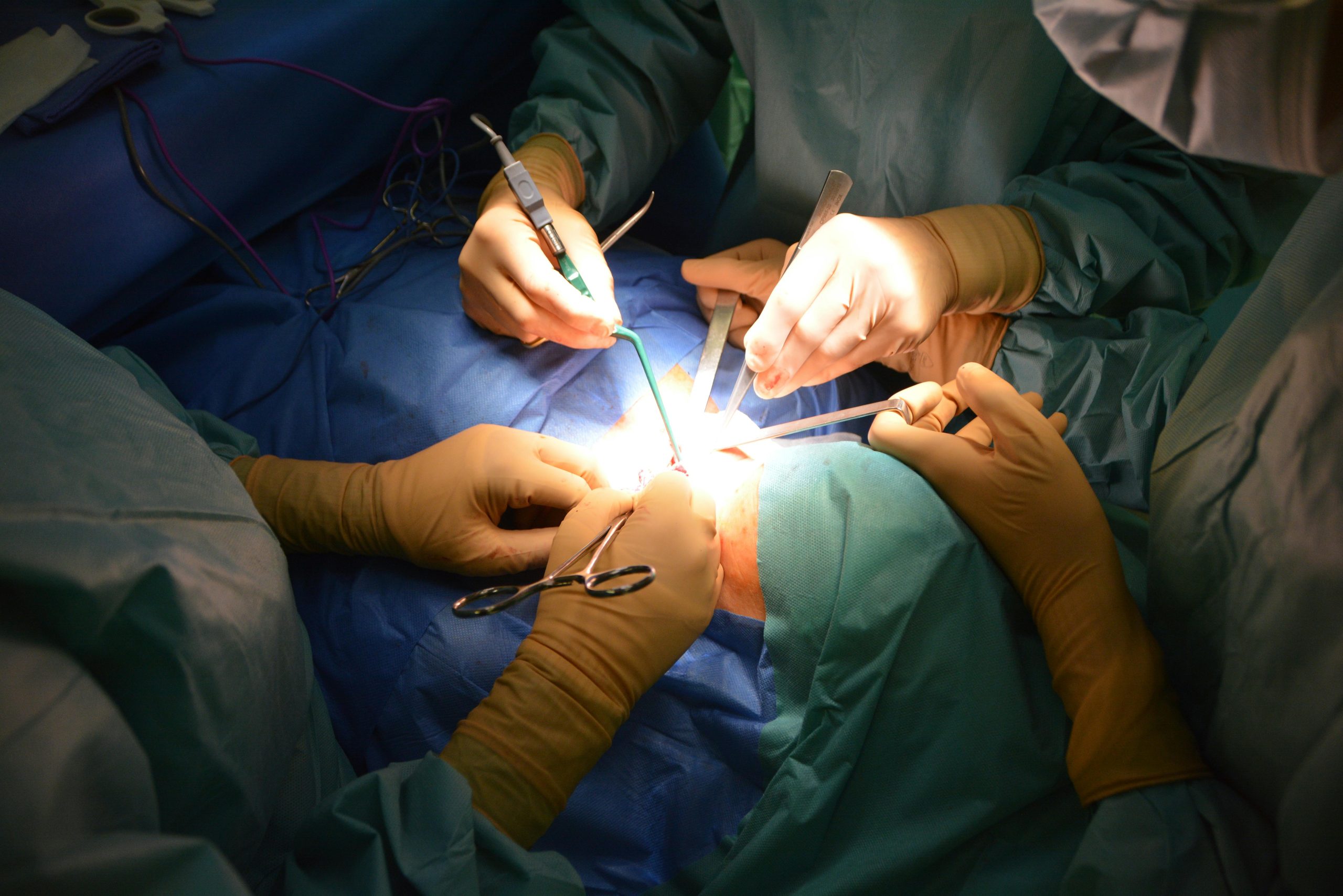
Surgical Options
Surgery may be necessary for complicated diverticulitis unresponsive to conservative measures, recurrent episodes significantly impacting quality of life, or fistula and stricture complications. Your doctor can discuss whether surgery might be appropriate based on your specific situation, frequency of episodes, and overall health status.
Emergency surgery for perforation with peritonitis may involve Hartmann’s procedure—the surgeon removes the diseased sigmoid and creates an end colostomy (an opening in your abdomen where stool passes into a bag), with potential reversal later. Laparoscopic lavage (a keyhole surgery procedure where the surgeon washes out the abdomen without removing any colon) is an option for purulent peritonitis in selected cases.
Elective surgery after recovery from acute episodes allows primary anastomosis (the surgeon removes the diseased section and reconnects the healthy ends of the colon) without colostomy in most patients. Laparoscopic sigmoid colectomy (keyhole surgery to remove the sigmoid colon through small incisions) offers faster recovery, less pain, and shorter hospitalisation compared to open surgery. The operation removes the diseased sigmoid segment whilst preserving the healthy colon and rectum.
Dietary Management for Prevention
Increasing Fibre Intake
Gradual fibre increase to recommended daily amounts can help reduce recurrence risk. Sudden large increases cause bloating and discomfort. Add fibre incrementally over several weeks. Soluble fibre (a type of fibre that dissolves in water and helps soften stool, found in oats, legumes, and fruits) and insoluble fibre (a type of fibre that adds bulk to stool, found in whole grains and vegetables) both contribute to stool bulk.
Psyllium husk supplements (a natural fibre derived from plant seeds) provide a convenient supplement when dietary sources are insufficient. Adequate fluid intake—at least several glasses daily—accompanies an increase in fibre to prevent constipation caused by inadequately hydrated fibre.
Foods to Emphasise
- Vegetables: broccoli, carrots, leafy greens, Brussels sprouts
- Fruits: berries, pears, apples with skin, oranges
- Legumes: lentils, chickpeas, black beans
- Whole grains: brown rice, quinoa, whole wheat bread
- Nuts and seeds (previously discouraged, now considered safe)
Eating Patterns
Regular meal timing promotes consistent colonic motility. Smaller, frequent meals rather than large infrequent ones reduce peak digestive demands. Thorough chewing improves initial breakdown and reduces colonic workload.
✅ Quick Tip
Add one new high-fibre food per week rather than overhauling your entire diet at once. This approach allows your gut bacteria to adapt gradually, minimising gas and bloating whilst building sustainable long-term habits.
When to Seek Professional Help
- Persistent abdominal pain lasting more than several hours, particularly if worsening
- Fever above normal levels with abdominal symptoms
- Inability to keep fluids down due to nausea or vomiting
- Significant change in bowel habits persisting beyond a few days
- Blood in stool or black, tarry stools
- Abdominal rigidity or severe tenderness to touch
- Symptoms not improving after a few days of prescribed treatment
- Recurrent episodes occurring frequently
Commonly Asked Questions
Can diverticulitis heal completely without surgery?
Uncomplicated diverticulitis typically resolves fully with conservative treatment. The acute inflammation heals, and normal digestive function returns. However, the diverticula themselves remain permanently, creating ongoing risk for future episodes. Surgery is reserved for complicated cases or frequent recurrences affecting quality of life.
How long does recovery from a diverticulitis episode take?
Response times vary depending on your specific condition. Uncomplicated cases usually improve within several days of starting treatment, with complete recovery over a few weeks. Complicated cases requiring hospitalisation or drainage take longer—often several weeks before resuming normal activities. Post-surgical recovery ranges from several weeks to longer, depending on the procedure type.
Is diverticulitis hereditary?
Genetic factors influence collagen structure and connective tissue strength, contributing to susceptibility. Having a first-degree relative with diverticular disease can increase risk. However, lifestyle factors—diet, exercise, weight—significantly modify this inherited predisposition and remain the primary targets for prevention.
Can I exercise with diverticulosis?
Regular physical activity is encouraged and can help reduce the risk of diverticulitis. Moderate exercise promotes healthy colonic motility and helps maintain appropriate body weight. During acute inflammation, rest is necessary, but exercise should resume once recovery is complete. Avoid heavy straining or Valsalva manoeuvres that increase intra-abdominal pressure.
Do I need regular monitoring after diverticulitis?
A colonoscopy several weeks after an acute episode may be recommended to exclude malignancy and assess for complications. Subsequent surveillance depends on findings and other risk factors. Patients with recurrent episodes or complications may benefit from closer follow-up with their colorectal surgeon to discuss preventive strategies, including possible elective surgery.
Conclusion
Diverticulitis develops when structural vulnerabilities in the colon wall combine with lifestyle factors such as low dietary fibre intake and sedentary behaviour. Adequate fibre intake and regular physical activity reduce recurrence risk. Prompt medical attention for persistent abdominal pain or fever prevents complications.
If you’re experiencing persistent left-sided abdominal pain, fever, changes in bowel habits, or recurrent diverticulitis episodes, consult a colorectal surgeon to evaluate appropriate treatment options.