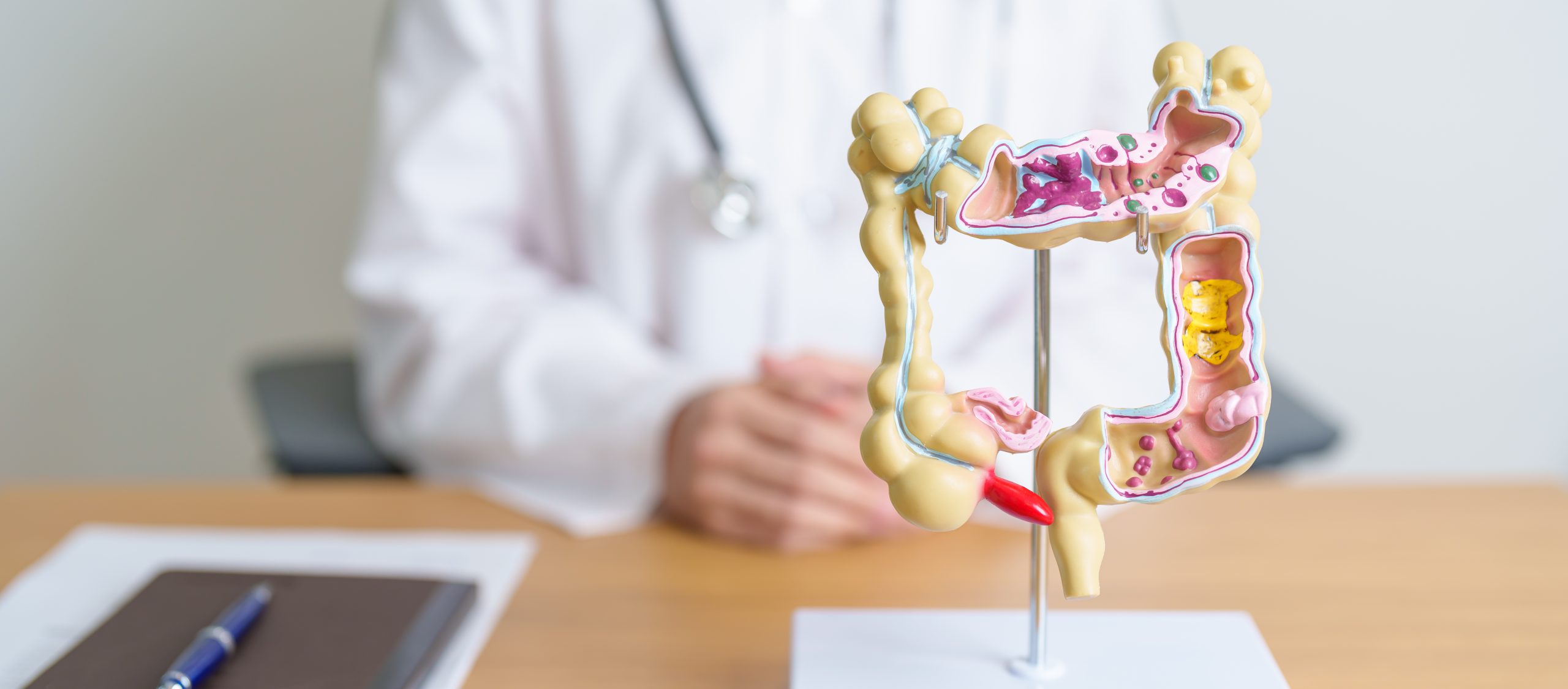
Colon polyps are abnormal tissue growths that develop on the inner lining of the colon or rectum. Most polyps remain harmless. However, certain types can transform into colorectal cancer over a period of years to decades through a well-documented biological process. This adenoma-to-carcinoma sequence makes polyp identification and removal a cancer prevention strategy in medicine. Colonoscopy screening (a procedure that uses a flexible tube with a camera to examine the inside of your colon) has become a component of preventive healthcare.
How Polyps Form in the Colon
The colon’s inner lining continuously regenerates, with cells dividing and replacing themselves in an orderly fashion. Polyps develop when genetic mutations disrupt this controlled cell turnover, causing abnormal cells to accumulate and protrude into the colon’s interior as visible growths ranging from a few millimetres to several centimetres.
- Genetic changes: The transformation from normal tissue to polyp involves multiple mutations that accumulate over time, progressing from a small adenoma toward increasingly abnormal cellular patterns — a multi-step process that typically spans years, creating a window for detection before cancer develops.
- Lifestyle and environment: Diets high in processed meats and low in fibre promote cellular mutations in the colon lining, while chronic inflammation from conditions like inflammatory bowel disease accelerates genetic damage.
- Age: Accumulated genetic changes become more likely with each decade of life, making age one of the strongest risk factors for polyp formation.
Types of Colon Polyps and Their Cancer Risk
Adenomatous Polyps
Adenomas represent the polyp type most associated with cancer development. These growths contain glandular tissue (cells that produce mucus or other substances) that displays abnormal cellular patterns under microscopic examination. Three subtypes exist:
- Tubular adenomas with tube-shaped glands
- Villous adenomas with finger-like projections
- Tubulovillous adenomas combining both patterns
Villous adenomas have a higher transformation risk than other adenoma subtypes. Their irregular surface architecture creates more opportunities for additional genetic mutations to occur. Size correlates directly with danger. Adenomas larger than 1 centimetre are more likely to harbour abnormal cellular changes than smaller growths.
Sessile Serrated Polyps
These flat or slightly raised polyps follow a different pathway to cancer than traditional adenomas. Their serrated, saw-toothed appearance under the microscope reflects distinct genetic mutations, particularly in genes that regulate cell growth. Sessile serrated polyps located in the right colon require careful surveillance due to their subtle appearance and transformation potential.
Hyperplastic Polyps
Most hyperplastic polyps pose minimal cancer risk. They are considered benign findings. These small, pale growths typically appear in the lower colon and rectum. However, larger hyperplastic polyps or those found in the right colon warrant closer attention. Distinguishing them from sessile serrated polyps can be challenging during colonoscopy.
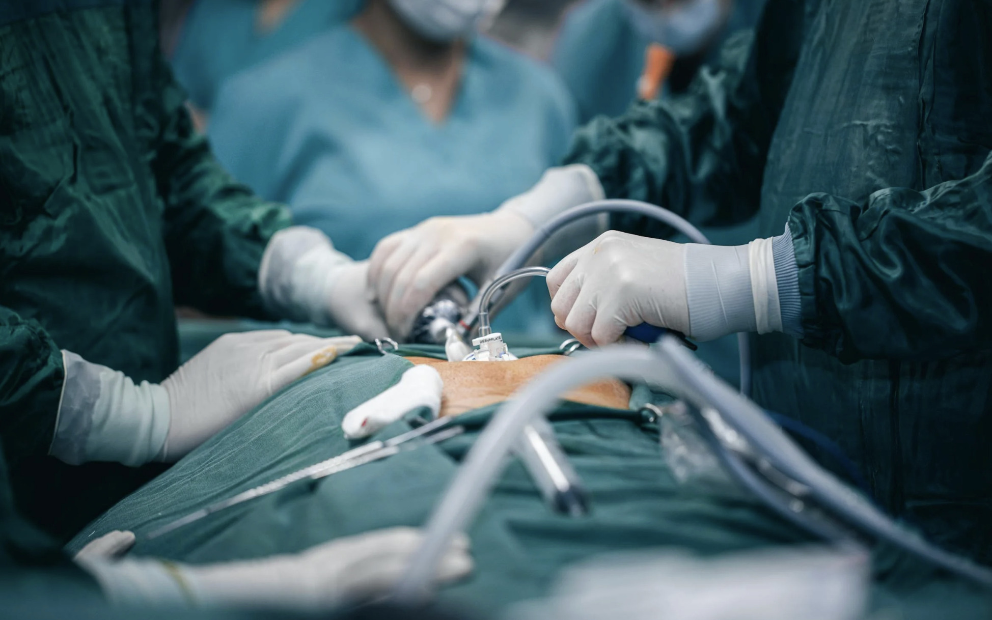
The Polypectomy Procedure
Polyp removal during colonoscopy (called polypectomy) eliminates precancerous tissue before transformation occurs. The technique used depends on polyp size, shape, and location within the colon.
Cold Snare Polypectomy
A thin wire loop encircles polyps smaller than one centimetre. It mechanically cuts through the base without an electrical current. This technique minimises thermal injury to surrounding tissue and reduces bleeding risk. Most small polyps encountered during routine screening undergo cold snare removal.
Hot Snare Polypectomy
Larger polyps require electrocautery (using electrical current to cut and seal tissue) to cut through thicker tissue stalks and seal blood vessels simultaneously. The snare delivers an electrical current while tightening around the polyp base. It cauterises as it cuts. This approach handles pedunculated polyps—those attached by a stalk—effectively.
Endoscopic Mucosal Resection
Flat or sessile polyps spreading across the colon surface require specialised removal techniques. A healthcare professional injects fluid beneath the polyp to lift it away from the underlying muscle layer. This creates a safety cushion. The elevated tissue is then removed in sections using snare techniques. This allows complete removal of larger lesions that would otherwise require surgery.
What Happens to Removed Polyps
Every polyp removed during a colonoscopy undergoes laboratory analysis. Pathologists (doctors who specialise in diagnosing disease by examining tissue samples) examine thin tissue sections under microscopes. They determine polyp type, assess cellular abnormality levels, and confirm complete removal.
Histological grading (the classification of how abnormal cells appear under a microscope) describes how abnormal the cells appear. Low-grade dysplasia (mild abnormal cell changes) indicates mild cellular changes. High-grade dysplasia (more severe abnormal cell changes) represents more pronounced abnormalities approaching early cancer. The pathology report guides subsequent surveillance recommendations.
Margin assessment confirms whether the polyp was completely removed. Clear margins indicate no abnormal tissue extends to the cut edge. This suggests successful removal. Positive or uncertain margins may prompt earlier follow-up colonoscopy or additional intervention.
Reports typically take several days to finalise. The findings, combined with polyp number, size, and location, determine personalised surveillance intervals for future colonoscopies. A healthcare professional will set surveillance schedules based on specific findings and risk factors.
Surveillance After Polyp Removal
Follow-up colonoscopy timing depends on initial findings. A colorectal surgeon will establish specific surveillance intervals tailored to individual risk factors. These include the number, size, and type of polyps found. Patients with 1 or 2 small tubular adenomas generally return after several years. Those with several adenomas, any adenoma larger than one centimetre, or adenomas with villous features or high-grade dysplasia typically require repeat examination within a few years.
Sessile serrated polyps larger than 1 centimetre or those with dysplasia warrant closer surveillance, often at 3-year intervals. Multiple serrated polyps may indicate serrated polyposis syndrome. This requires annual surveillance and evaluation of family members.
Incomplete polyp removal necessitates short-interval follow-up, usually within several months. This allows examination of the removal site and addresses any residual tissue. Clear subsequent examinations allow return to standard surveillance intervals.
Factors Affecting Polyp Recurrence
Previous polyp findings predict future polyp development. Patients who have had adenomas removed are more likely to develop additional adenomas compared to those with negative colonoscopies. The number and characteristics of initial polyps correlate with recurrence rates.
Family history influences recurrence patterns. First-degree relatives with colorectal cancer or adenomas indicate shared genetic susceptibility. This elevates personal risk for ongoing polyp formation.
Modifiable factors play a role in recurrence:
- Maintaining a healthy weight
- Engaging in regular physical activity
- Limiting red and processed meat consumption
- Avoiding tobacco use
These lifestyle factors create conditions less favourable to polyp development. They complement but do not replace surveillance colonoscopy.
Recognising Symptoms That Warrant Evaluation
Most colon polyps produce no symptoms. They grow silently until discovered during screening. However, larger polyps occasionally cause noticeable changes that prompt medical evaluation outside routine screening schedules.
- Rectal bleeding appearing as bright red blood on toilet paper or in the bowl may indicate polyps. However, haemorrhoids and other conditions cause similar symptoms.
- Changes in bowel habits (such as new constipation, diarrhoea, or narrowed stools) persisting beyond several weeks warrant investigation.
- Iron deficiency anaemia (low red blood cell count due to insufficient iron) without an obvious cause can result from slow, chronic bleeding from large polyps.
Abdominal discomfort rarely results solely from polyps. However, large growths occasionally cause vague cramping or fullness. Any combination of these symptoms, particularly in individuals over a certain age or with a family history of colorectal conditions, merits medical consultation.
Commonly Asked Questions
How long does it take for a polyp to become cancerous?
The adenoma-to-carcinoma progression typically spans a number of years. However, response times vary depending on specific conditions. Aggressive subtypes or those with high-grade dysplasia may progress faster. This extended timeframe explains why screening at recommended intervals effectively intercepts most polyps before transformation occurs.
Will I feel anything during polyp removal?
Colonoscopy sedation ensures comfort throughout the procedure, including during polyp removal. The colon lining lacks pain receptors. Polypectomy produces no sensation. Post-procedure, mild bloating from air insufflation resolves within hours. Larger polypectomies occasionally cause minor cramping that settles quickly.
Can polyps grow back after removal?
Completely removed polyps do not regrow at the same site. However, patients who develop polyps have demonstrated susceptibility to polyp formation. They commonly develop new polyps elsewhere in the colon over subsequent years. This pattern underlies the need for surveillance colonoscopy following initial polyp detection.
How do I prepare for a colonoscopy?
Bowel preparation involves the following steps:
- Follow a clear liquid diet the day before
- Drink a prescribed laxative solution that cleanses the colon
Thorough preparation improves polyp detection rates by providing clear visualisation of the colon lining. Specific instructions vary between preparation products. A healthcare professional provides detailed guidance before the procedure.
Does polyp removal guarantee I won’t get colorectal cancer?
Removing polyps substantially reduces colorectal cancer risk by eliminating precancerous tissue. However, new polyps can develop between surveillance intervals. Rarely, cancers arise from flat lesions that are difficult to detect. Maintaining recommended surveillance schedules and reporting new symptoms provides ongoing protection.
Next Steps
Polyp removal interrupts cancer development before transformation occurs. Pathology results determine surveillance intervals. Maintaining recommended colonoscopy schedules and reporting new symptoms maximises cancer prevention.
If you’re experiencing rectal bleeding, changes in bowel habits, or unexplained anaemia, consult a qualified colorectal surgeon to discuss appropriate screening or treatment options tailored to your individual circumstances.