

- Rectal Cancer: TaTME is primarily performed for patients with middle to low rectal cancer, typically located within 12 cm from the anal verge. The technique allows for precise dissection in areas difficult to access through conventional approaches.
- Anatomical Constraints: Patients with a narrow pelvis, high body mass index, or bulky tumours benefit from TaTME as these anatomical features can limit access and visibility during traditional surgical approaches.
- Previous Pelvic Surgery: Patients who have undergone previous pelvic operations may have scarring and adhesions that make conventional surgery more difficult. TaTME provides an alternative approach that can navigate through scarred tissue.
- Need for Sphincter Preservation: This approach may be selected when preservation of the anal sphincter function is a priority and the tumour is located close to the sphincter complex.
- Following Neoadjuvant Therapy: Patients who have received chemotherapy and/or radiotherapy before surgery (neoadjuvant therapy) and show good response may be candidates for TaTME as part of a sphincter-sparing strategy.
- Bail out for complications encountered from traditional approaches: During surgery via traditional approaches, if bleeding is encountered and visualisation is lost, TaTME may be able to complete the surgery from the bottom-up approach thereby averting further delay in arresting the bleeding or need for conversion.
TransAnal Total Mesorectal Excision (TaTME) Treatment in Singapore
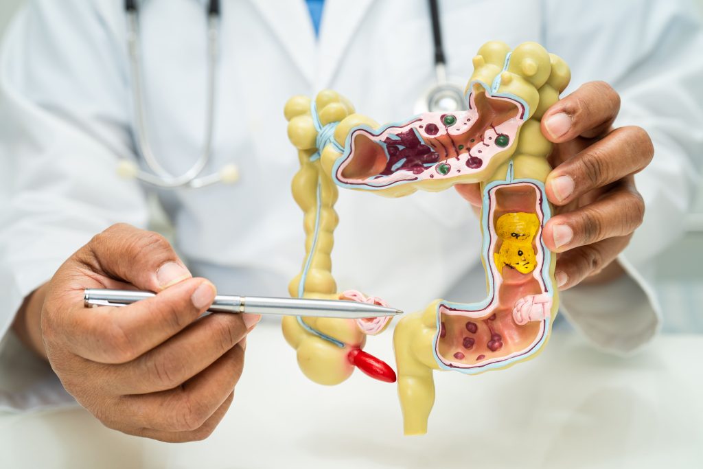
TransAnal Total Mesorectal Excision (TaTME) is a surgical procedure that removes the rectum and surrounding tissues (mesorectum) through a combined approach using both abdominal and transanal access points. This technique addresses rectal cancer by extracting the affected section of the bowel along with lymph nodes that may contain cancer cells. TaTME provides surgeons with better visualisation and access to the lower rectum, particularly in cases where anatomical constraints like a narrow pelvis make traditional approaches challenging.
Indications for TaTME
The following conditions and situations may determine whether TaTME would be an appropriate surgical approach for a patient’s specific case.
Benefits of TaTME
TaTME offers several advantages over conventional surgical approaches for treating rectal cancer and other rectal conditions.
Improved Visibility
The bottom-up approach allows surgeons to directly visualise the distal resection margin, enabling more controlled dissection in the deep pelvis where visibility is often limited in traditional approaches.
Better Access to Difficult Areas
TaTME provides improved access to the distal rectum, which can be challenging to reach adequately with conventional laparoscopic or open surgical methods.
CT Scan
This imaging technique provides detailed information about the location and cause of the obstruction. It also detects complications such as reduced blood supply (ischemia) or intestinal perforation, helping to determine whether emergency intervention is required.
Potentially Lower Positive Margin Rates
The technique may contribute to reduced rates of positive circumferential resection margins, which is a factor in reducing local recurrence of rectal cancer.
Sphincter Preservation
For appropriately selected patients, TaTME may increase the likelihood of preserving the anal sphincter while still achieving complete tumour removal.
Single Stapling Technique
TaTME allows for a single distal stapling technique, which may reduce the risk of anastomotic failure associated with multiple stapling techniques used in conventional approaches.
Surgical Techniques
Two-Team Simultaneous Approach
This is the currently most standard way of performing this surgery internationally. This method involves two surgical teams working concurrently, with one team performing the abdominal portion and the other the transanal portion of the procedure. The simultaneous approach reduces overall operative time and allows for coordinated dissection from above and below. The teams meet at a point in the pelvis, typically around the peritoneal reflection, to complete the mesorectal excision.
Sequential Approach
In this technique, the surgical team first completes the abdominal mobilisation of the colon and proximal rectum before proceeding to the transanal phase. The sequential method allows for systematic progression through the procedure but may result in longer operative times compared to the simultaneous approach. This is hardly done now unless there is a limitation in manpower.
Pure Transanal Approach
Some selected cases may be managed with a predominantly transanal approach, with minimal abdominal assistance. This technique is typically reserved for early-stage tumours in the distal rectum. The pure transanal approach involves creating a platform for the transanal access, performing the distal rectal dissection, and then continuing proximally to complete the total mesorectal excision.
Robotic-Assisted TaTME
This technique incorporates robotic platforms to enhance precision and control during the procedure. Robotic assistance can be utilised for either the abdominal component, the transanal component, or both phases of the operation. The robotic system provides improved dexterity and three-dimensional visualisation, which may be beneficial for complex dissections in the confined pelvic space.
Preparing for Surgery
- Medical Evaluation: A comprehensive assessment including a medical history review, physical examination, and diagnostic tests is conducted. This evaluation includes a colonoscopy with biopsy, an MRI of the pelvis, a CT scan of the chest and abdomen, and blood tests to check organ function and blood counts. This information is used to determine the stage of cancer and create a personalised surgical plan.
- Bowel Preparation: Cleansing of the bowel is typically required before surgery. This process involves dietary modifications starting several days before surgery, typically progressing from a low-residue diet to clear liquids. Patients must take prescribed laxatives or bowel cleansing solutions the day before surgery to ensure the colon and rectum are as empty as possible, reducing the risk of infection.
- Medication Adjustments: Certain medications may need to be stopped temporarily before surgery. Blood thinners such as aspirin, warfarin, or clopidogrel may need to be discontinued several days to a week before surgery to reduce bleeding risk.
- Pre-operative Fasting: Food and fluid intake must be restricted before surgery to reduce the risk of aspiration during anaesthesia. Typically, patients should not eat solid food for 6-8 hours before surgery and should avoid clear liquids for 2-4 hours before the procedure.
Step-by-Step Procedure
Anaesthesia Administration
The procedure begins with the administration of general anaesthesia by an anaesthesiologist. The patient is positioned, typically in a modified lithotomy position with slight Trendelenburg tilt, to optimise access to both the abdomen and perineum. Monitoring equipment is attached to track vital signs throughout the procedure.
Abdominal Phase Setup
The abdominal component usually begins with the creation of pneumoperitoneum and placement of laparoscopic ports. The surgeon examines the abdominal cavity to confirm the absence of metastatic disease. The inferior mesenteric vessels are identified and ligated, and the left colon is mobilised to provide adequate length for later reconstruction.
Transanal Phase Preparation
A specialised transanal platform is inserted through the anus and secured in place. Carbon dioxide is insufflated to create a working space in the rectum. The surgeon then marks the distal resection margin and performs a full-thickness circumferential rectal wall incision below the tumour.
Distal to Proximal Dissection
The mesorectal plane is identified and followed upward through careful dissection. The surgeon maintains the integrity of the mesorectal fascia, which is critical for oncological outcomes. The dissection continues in the avascular plane between the mesorectum and surrounding structures, gradually working upward toward the peritoneal reflection.
Meeting of Dissection Planes
The transanal dissection continues until it meets the abdominal dissection plane, typically at the level of the peritoneal reflection. This juncture completes the circumferential mobilisation of the rectum with its intact mesorectal envelope. The specimen remains attached only by the proximal bowel.
Specimen Extraction
After complete mobilisation, the specimen is extracted, either through an abdominal incision or transanally, depending on the size. The surgeon examines the specimen to ensure adequate resection margins and complete mesorectal excision before sending it for pathological assessment.
Reconstruction Phase
The continuity of the digestive tract is restored, typically by creating a colorectal or coloanal anastomosis. This connection may be performed using stapling devices or hand-sewn techniques, depending on the level of resection. In some cases, a temporary diverting stoma may be created to protect the anastomosis during healing.
Closure
The procedure concludes with inspection for haemostasis and irrigation of the pelvic cavity. Drains may be placed near the anastomosis to monitor for leakage. Laparoscopic port sites and any additional incisions are closed in layers, and dressings are applied.
Post-Surgical Care and Recovery
Immediate Care
After surgery, patients are closely monitored in a recovery area for vital signs, pain levels, and the condition of the surgical site. Pain management typically starts with intravenous analgesics and transitions to oral medications. Within 24 hours, patients are encouraged to begin moving to help prevent complications such as blood clots and pneumonia.
Diet and Stoma Care
Initially, patients are given clear liquids, and the diet is gradually advanced as bowel function returns. This progression goes from liquids to soft foods and eventually to a regular diet. For those with a stoma, specialised nurses provide education on proper stoma care and dietary adjustments to ensure optimal healing and comfort.
Wound Care and Functional Recovery
Surgical wounds are inspected daily for signs of infection, with dressings changed according to hospital protocols. Most patients can return to light activities within 2-4 weeks, while more strenuous activities are generally delayed for 6-8 weeks. If a temporary stoma was created, a reversal procedure is typically performed 8-12 weeks post-surgery once the primary surgical site has healed.
Follow-up
Regular follow-up visits are scheduled to monitor recovery and detect any potential cancer recurrence. These appointments often include physical exams, blood tests, and imaging studies. Changes in bowel function, such as increased frequency or urgency, are common but tend to improve over the course of 6-12 months as the body adjusts to the surgical changes.
Are Your Symptoms Affecting Your Quality of Life?
Consult our MOH-accredited specialist for an accurate diagnosis & personalised treatment plan today.

Potential Risks and Complications
TaTME carries risks common to major abdominal and pelvic surgeries, including infection, bleeding, and anastomotic leakage. Urinary and sexual dysfunction may occur due to nerve damage, and injury to nearby structures, such as the ureters or bladder, is rare but possible. Additional risks include pelvic sepsis, pelvic floor dysfunction, local cancer recurrence, and long-term bowel issues like frequency, urgency, or incontinence.
Frequently Asked Questions
How often will I need follow-up appointments after TaTME surgery?
Follow-up appointments after TaTME generally begin 2-4 weeks after the surgery, with subsequent visits spaced out every 3-6 months during the first 2-3 years. Over time, as the patient’s condition stabilises, the frequency of visits may decrease. The timing of follow-ups may vary based on individual recovery progress and the patient’s risk of cancer recurrence.
Is there a risk of cancer recurrence after TaTME surgery?
While TaTME offers high precision in removing rectal cancer, there is still a possibility of recurrence, especially if cancer cells were present in the resection margins. Regular follow-up visits and imaging studies help detect recurrence early, allowing for timely intervention if necessary.
What should I do if I experience changes in bowel function after surgery?
It is common to experience changes in bowel habits after TaTME, such as increased frequency, urgency, or incontinence. These issues usually improve over time, but if they persist or worsen, further treatments may be recommended to manage symptoms and improve quality of life.
Partnered Programmes & Insurance Plans
For Singaporeans, Singapore Permanent Residents and Foreigners. Please speak to our friendly clinic staff about using your insurance plans.


contact us
Please leave us a message and our friendly clinic staff will get back to you as soon as possible. For urgent or same day appointments, kindly call the clinic to arrange an appointment.
Our Clinic Locations
Ark Surgical Practice – Mount Elizabeth Medical Centre
3 Mount Elizabeth, #09-07
Singapore 228510
Monday to Friday: 9am – 5pm
Saturday: 9am – 12:30pm
Sunday & Public Holidays: Closed
Ark Surgical Practice – Mount Alvernia Hospital
820 Thomson Road,
Mount Alvernia Hospital, #06-52,
Medical Centre D, Singapore 574623
Wednesday: 9am – 12:30pm
Thursday: 2pm – 5pm